Nuclear research – an early oncology alert
Nuclear medicine techniques enable cancer diagnosis at the earliest stage – often when other diagnostic methods fail. With early diagnosis using PET-CT and SPECT-CT procedures, a patient’s chances of a complete cure or a significant improvement in prognosis are very high, and the costs of the entire diagnostic and therapeutic process are optimized, says Dr. med. Rafal Czepczynski of the Polish Society of Nuclear Medicine.
In 2019, oncological diseases invariably remain one of the most common causes of deathoin the world. The incidence is increasing year after year, and the morbidity forecasts are so alarming that experts mobind o „oncology tsunami”. Although new cancer prevention approaches and new therapies are being implemented into clinical practice, there is no single fully effective cure for cancer.
Medicaid and public health experts stress that oprocz attention to a healthy lifestyle – primarily a daily dose of exercise and a healthy diet – key to effectiveness in the fight against cancer, the study found – zarowno these basic, prophylactic, as well as specialized, ktore are particularolnie important when suspicion of cancer is highly likely and at the stage of oncological treatment, when a possible recurrence of the disease can be detected early enough that the condition is often fully treatable.
– As physicians in our daily practice, we see the importance of early cancer diagnosis – bothoboth for the physical well-being and health of the patient, as well as for his mental state and comfort in therapy. There is no doubt that rapid and accurate testing significantly increases the chances of success in the fight against cancer – It is worth using them for the benefit of the patientoin and for the state budget – The cost of the diagnostic and therapeutic process and also patient care and rehabilitationow are significant and growing expenses. It has been emphasized for years that every country in the world must optimize these – Poland also. In the field of cancer diagnosis and therapyow procedures from the field of nuclear medicine are helpful – precise, fast and non-invasive, as comfortable as possible for the patient – explains dr hab. med. Rafal Czepczynski of the Polish Society of Nuclear Medicine.
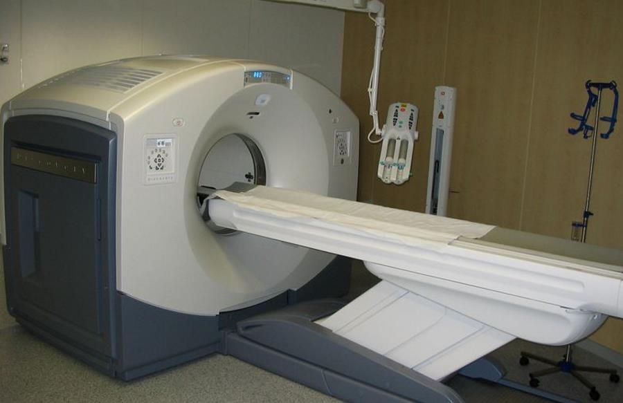
Cancer diagnostic methods include roA variety of procedures, used depending, among other things, on the type of cancer suspected, among theod them: histopathological examinations, immunohistochemical procedures, blood count, biochemical tests, in the case of tumorow with a genetic basis genetic and molecular diagnostic procedures, as well as imaging procedures: x-ray, mammography, computed tomography, magnetic resonance imaging, positron emission tomography (PET) and single photon emission tomographyow (SPECT). During examinations: PET and SPECT, simultaneous computed tomography (CT) can be performed – woat the time of the study will be determined as PET-CT and SPECT-CT.
Positron emission tomography study – PET. Positron emission tomography) involves the registration of radiation resulting from positron emissionow by isotope-labeled medical preparations – radiopharmaceuticals.
– Positron emission tomography testing is used, among other things, in patientsoin whomor who have been diagnosed with neoplasmor malignant, and with other diagnostic methods it is not possible to determine where the primary focus of the disease is located. PET is also performed when it is necessary to assess how extensive cancerous lesions are – before deciding on the best therapy to implement. PET examination is very sensitive, allows early diagnosis of malignant lesions, including small metastasesow. With PET, we can determine whether there has been a recurrence after surgical treatment and whether the treatment used, such as chemotherapy, is having a good effect – explains dr hab. med. Rafal Czepczynski.
Single photon emission tomographyow -SPECT. Single photon emission computer tomography (PET) is a similar, non-invasive imaging test to PET, using isotopes of radiotworcze. In this case for imaging the organogamma emitting radiopharmaceuticals are used.
– SPECT technique is an excellent addition to the diagnostic palette of cancerow, among other scintigraphic examination, as it allows imaging of lesions in 3D. We can pinpoint disease foci very precisely and detect lesions smaller than with scintigraphy – It is crucial to the diagnostic success of. As nuclear medicine specialists, we appreciate the Ministry of Health’s funding for the facilityoIn nuclear medicine, whichore helped to purchase modern diagnostic equipment. These investments should be followed by a steady increase in the availability of nuclear medicine procedures – The needs in this area are constantly growing – mowi dr hab. med. Rafal Czepczynsk.


